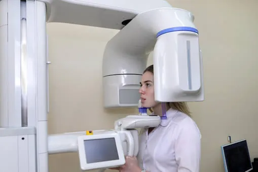
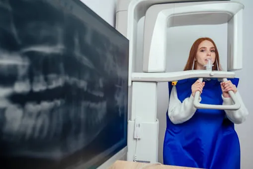
What is Cone Beam CT 3D Imaging?
Cone beam computed tomography (CT) is a specialized x-ray machine used when regular dental or facial x-rays are insufficient. While not used routinely due to increased radiation exposure compared to regular dental x-rays, cone beam CT generates highly detailed 3-D images of dental structures, soft tissues, nerve paths, and bone in the craniofacial region in a single scan. This allows for precise treatment planning, similar to conventional CT imaging. Unlike conventional CT, dental cone beam CT uses a smaller, less expensive machine that can be placed in an outpatient office. While cone beam CT provides detailed images of the bone, it is not as effective as conventional CT in evaluating soft tissue structures such as muscles, lymph nodes, glands, and nerves. However, it does offer lower radiation exposure, making it a safer option for patients.
What are Some Common Uses of the Procedure?
Dental cone beam CT is commonly employed to aid in the treatment planning of various dental issues. It is especially useful in more complex cases that involve:
- Surgical planning for impacted teeth
- Diagnosis of temporomandibular joint disorder (TMJ)
- Accurate placement of dental implants
- Evaluation of the jaw, sinuses, nerve canals, and nasal cavity
- Detection, measurement, and treatment of jaw tumors
- Determination of bone structure and tooth orientation
- Identification of the origin of pain or pathology
- Cephalometric analysis
- Reconstructive surgery
How Should I Prepare for a Cone Beam CT in Sunrise FL?
Preparation for a cone beam CT examination is simple and straightforward.
Before the procedure, you will need to remove any metal objects that may interfere with the imaging, including jewelry, eyeglasses, hairpins, and hearing aids. While removable dental work may also need to be taken out, it is recommended to bring them to your examination as your dentist or oral surgeon may need to examine them.
For female patients, it’s crucial to inform your dentist or oral surgeon if there is any chance that you might be pregnant. You can refer to the Safety page for more information about the risks of x-rays during pregnancy. Otherwise, there is no special preparation required for the examination.
What Are the Advantages Vs. Risks?
Advantages:
- Cone beam CT produces high-quality images due to the focused x-ray beam, reducing scatter radiation.
- A single scan provides a comprehensive evaluation of the area of interest, with multiple views and angles that can be manipulated.
- Cone beam CT scans provide more precise treatment planning compared to conventional dental x-rays.
- CT scanning is accurate, noninvasive, and painless.
- CT scanning can simultaneously image bone and soft tissue.
- There is no residual radiation left in the body after a CT exam.
Risks:
- Although the immediate side effects of the x-rays used in CT scanning are negligible, there is always a slight risk of cancer from excessive radiation exposure. However, the benefits of an accurate diagnosis usually outweigh the risks.
- Children are more sensitive to radiation and should only have CT exams if they are necessary for a diagnosis. Repeated CT exams should be avoided, and low-dose techniques should be used for CT scans in children.